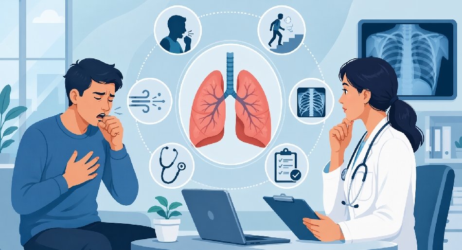
Breathing is an autonomic process, a continuous cycle governed by the brainstem that occurs roughly 20,000 times a day. For most, this requires zero conscious effort. However, when the mechanics of respiration become noticeable or labored, it serves as a primary clinical indicator of physiological stress. Patients often attempt to rationalize a decline in lung function, attributing a lingering cough to seasonal changes or dismissing breathlessness as a byproduct of aging.
In a clinical setting, distinguishing between a self-limiting respiratory event and a progressive pulmonary pathology is essential for long-term outcomes. While primary care providers manage the bulk of acute respiratory infections, persistent symptoms require a transition to specialized care. Determining when to involve a pulmonologist involves a rigorous assessment of symptom duration, severity, and the failure of first-line interventions.
The Physiology of the Persistent Cough
A cough is a protective reflex designed to clear the airways of mucus, irritants, or foreign bodies. It involves a coordinated sequence: an inspiratory phase, a forced expiratory effort against a closed glottis, and the sudden opening of the glottis to release air at high velocity. While effective for clearing the occasional irritant, a cough that becomes chronic indicates a state of persistent inflammation or hypersensitivity within the respiratory tract.
The medical community generally follows a duration-based classification system for coughs:
- Acute: Lasting less than three weeks (usually viral or bacterial).
- Subacute: Lasting three to eight weeks (often post-infectious).
- Chronic: Persisting beyond eight weeksCHEST Guideline.
When a cough reaches the eight-week mark, the likelihood of it resolving without targeted intervention decreases significantly. At this stage, the diagnostic focus shifts from treating a suspected infection to identifying structural or systemic causes.
The Diagnostic “Triple Threat”
In over 90% of non-smoking patients with a normal chest X-ray, chronic cough is caused by one or more of three conditions:
- Upper Airway Cough Syndrome (UACS): Formally known as postnasal drip, where secretions from the sino-nasal cavity irritate the pharyngeal cough receptors.
- Asthma: Specifically, cough-variant asthma, where the classic wheeze is absent, but bronchial hyperresponsiveness remains.
- Gastroesophageal Reflux Disease (GERD): Acid or non-acid reflux can trigger the cough reflex either through micro-aspiration into the larynx or via a vagally mediated sensory reflex from the distal esophagus.
If a patient fails to respond to empirical trials for these conditions—such as nasal steroids, bronchodilators, or proton pump inhibitors—the involvement of a pulmonologist is necessary to rule out more complex etiologies like bronchiectasis or interstitial lung disease.
Clinical Triage of Respiratory Symptoms
| Symptom | Typical/Benign Presentation | Specialist Referral Required |
|---|---|---|
| Cough Duration | Resolves within 21 days post-infection. | Persists beyond 8 weeks (Chronic). |
| Sputum Quality | Clear, thin, or white. | Hemoptysis (blood-tinged), rust-colored, or foul-smelling. |
| Dyspnea | Occurs during high-intensity exertion. | Occurs during ADLs (walking, dressing) or at rest. |
| Chest Sensation | Generalized soreness from coughing. | Sharp pleuritic pain, localized tightness, or stridor. |
| Oxygen Saturation | Stable at 95%–100% on room air. | Frequent dips below 92%ATS Consensus or sustained hypoxia. |
Evaluating Dyspnea: When Breathlessness Signals Pathology
Dyspnea, or the subjective sensation of breathing discomfort, is a complex symptom involving sensory input from multiple receptors in the lungs, upper airways, and chemoreceptors in the brain. It is often described by patients as “air hunger,” “chest tightness,” or “increased effort to breathe.”
While exertional dyspnea is normal at the limits of physical capability, it becomes pathological when it occurs during activities that were previously tolerated. A pulmonologist uses various scales, such as the Modified Medical Research Council (mMRC) Dyspnea Scale, to quantify this impairment.
Shortness of breath is rarely an isolated issue. Because the heart and lungs operate as a single functional unit, pulmonary insufficiency often mirrors cardiac failure. A pulmonologist’s role is to determine if the dyspnea is obstructive (difficulty getting air out, as in COPD), restrictive (difficulty getting air in, as in pulmonary fibrosis), or a result of pulmonary vascular disease (problems with blood flow through the lungs).
Seven Clinical Triggers for a Pulmonary Referral
The following conditions and symptoms warrant a specialist’s evaluation to prevent irreversible damage to the lung parenchyma or vasculature.
1. The Refractory Chronic Cough
As discussed, a cough lasting more than 8 weeks is not normal. A pulmonologist can perform advanced testing, such as methacholine challenge tests to identify occult asthma or bronchoscopy to visualize the internal airway architecture if the cause remains elusive after standard imaging. For related guidance, see [Pulmonary Function Tests](/blog/pulmonary-function-tests-what-they-are-why-you-might-need-one/).
2. Hemoptysis (Coughing Up Blood)
The expectoration of blood from the lower respiratory tract is a high-priority clinical sign. Whether it appears as minor streaks or significant volume, it mandates an immediate investigation. Causes range from acute bronchitis to more severe pathologies like pulmonary embolism, tuberculosis, or bronchogenic carcinoma.
3. Progressive Exertional Dyspnea
When a patient reports they can no longer walk a block or climb stairs without stopping, it indicates a significant reduction in pulmonary reserve. This requires a full workup to differentiate between airway disease, interstitial disease, or pulmonary hypertension.
4. New or Persistent Wheezing
Wheezing is a high-pitched whistling sound produced by air moving through narrowed small airways. While often associated with asthma, new-onset wheezing in an adult can also signal localized obstruction, such as a tumor or foreign body, or the onset of Chronic Obstructive Pulmonary Disease (COPD).
5. Abnormal Radiographic Findings
Incidental findings on a chest X-ray or CT scan—such as lung nodules, hilar lymphadenopathy, or “ground-glass” opacities—require expert interpretation. A pulmonologist manages the longitudinal tracking of these findings or coordinates biopsies to rule out malignancy or sarcoidosis.
6. Hypoxemia and Chronic Fatigue
Low blood oxygen levels (hypoxemia), even if asymptomatic at rest, can lead to chronic fatigue, cognitive “fog,” and eventual right-sided heart failure (cor pulmonale). If a patient’s pulse oximetry consistently reads below 92%ATS Consensus, a specialist must determine the cause of the gas exchange impairment.
7. Occupational or Environmental Exposures
Patients with a history of working in construction, mining, manufacturing, or firefighting are at increased risk for occupational lung diseases. Exposure to silica, asbestos, coal dust, or toxic fumes can lead to delayed-onset fibrosis or pleural disease that requires specialized monitoring.
The Intersection of Respiratory Health and Sleep
Pulmonary medicine and sleep medicine are deeply intertwined. Many respiratory pathologies are exacerbated during sleep due to changes in thoracic mechanics and a decrease in central respiratory drive.
Obstructive Sleep Apnea (OSA) is perhaps the most prevalent condition where the upper airway collapses during sleep, leading to repetitive cycles of hypoxia and arousal. However, pulmonologists also manage “Overlap Syndrome,” where a patient has both COPD and OSA. This combination significantly raises cardiac risk — see [Overlap Syndrome details](/blog/shift-work-sleep-disorder-when-your-schedule-turns-sleep-into-a-struggle/). This combination leads to more profound nocturnal desaturation than either condition alone, significantly increasing the risk of cardiac arrhythmias and pulmonary hypertension.
Effective management of sleep-disordered breathing goes beyond simple snoring cessation; it is about protecting the cardiovascular system from the repetitive stress of low oxygen. Specialized interventions, including CPAP titration and airway optimization, are fundamental components of pulmonary care. American Lung Association: When to See a Pulmonologist
Diagnostic Procedures in the Pulmonary Clinic
When a patient visits a specialist, the diagnostic approach is more granular than in primary care.
Pulmonary Function Testing (PFT)
PFTs are the gold standard for assessing lung health. They involve:
- Spirometry: Measuring how much and how fast air can be exhaled.
- Lung Volumes: Determining the total capacity of the lungs, including the air left over after a full exhale (residual volume).
- Diffusion Capacity (DLCO): Measuring how well oxygen moves from the lung air sacs into the blood. This is a critical metric for detecting damage to the alveolar-capillary membrane.
Imaging and Bronchoscopy
High-Resolution Computed Tomography (HRCT) provides a detailed view of the lung’s “skeleton” (the interstitium). If imaging is inconclusive, a pulmonologist may perform a bronchoscopy—inserting a thin, flexible camera into the airways—to take tissue samples or perform a bronchoalveolar lavage (sampling the fluid in the lungs).
Management Strategies for Chronic Lung Disease
The goal of pulmonary intervention is twofold: symptom mitigation and the preservation of existing lung function. Since lung tissue generally does not regenerate, early intervention is the only way to halt the progression of chronic diseases.
Pharmacological Interventions
Treatment often involves a combination of:
- Long-Acting Bronchodilators: These keep the airways open for 12 to 24 hours.
- Inhaled Corticosteroids: Targeted anti-inflammatory agents that reduce airway swelling with minimal systemic side effects.
- Biologics: For severe, eosinophilic asthma, monoclonal antibodies can target specific inflammatory pathways.
Pulmonary Rehabilitation
Rehabilitation is a highly effective, non-pharmacological intervention. It involves a structured program of exercise, breathing retraining (such as pursed-lip breathing), and nutritional support. The objective is to increase the efficiency of the skeletal muscles, allowing the patient to do more with less respiratory effort.